Hipofisis


Human pituitary stained using the 'Orange G/Acid fuchsin/Light green' technique. Formalin-fixed, paraffin embedded tissue, 4µm thick section.

Result: Acidophils - orange/yellow
Basophils - magenta red
Chromophobes - grey/green
© Roy C. Ellis 2004Basophils - magenta red
Chromophobes - grey/green
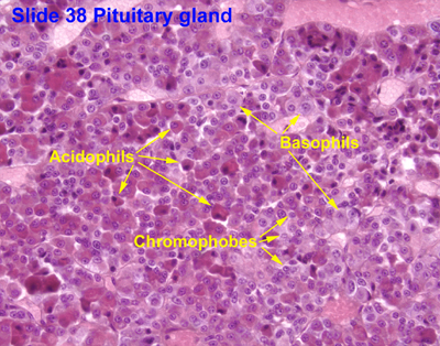
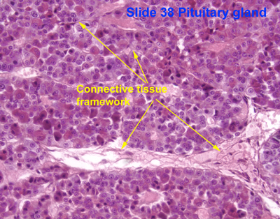
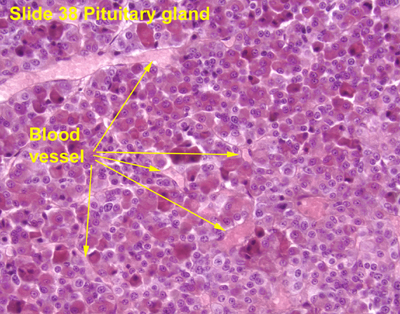
ø PARS DISTALIS
1. Cromofobas
- Células cormófilas degranudadas. Son las anteriores, que han liberado por exocitosis sus gránulos. También puede haber indiferenciadas.
- Células folículo-estrelladas. No son secretoras. Pueden adquirir dos formas:
· Células con muchas prolongaciones o estrelladas (estrelladas).
˙ Granulos de glucogeno
2. Cromofilas:
¬ Acidofilas
Nucleo excentrico y redondeado.
≈ Somatotropas:are cells in the anterior pituitary that produce
1. growth hormone. These cells constitute 30-40% of anterior pituitary cells.
+They respond by releasing GH in response to Growth hormone releasing hormone (GHRH, or somatocrinin)
-are inhibited by GHIH(somatostatin), both received from the hypothalamus via the hypophyseal portal system vein and the secondary plexus.
Somatotrope cells are classified as acidophilic cells. These cells take years to grow and mature very slowly. If these cells grow large enough they can impair vision, cause headaches or damage other pituitary functions.
These cells produce growth hormone in response to GHRH. If there is an excess of growth hormone it is usually because of over-secretion of somatotrope cells in the anterior pituitary gland. This malfunction characterizes the disease gigantism.
-SOMATOTROPINA
-GROWTH H.
cerca de capilares
≈ Lactotropas: are cells in the anterior pituitary which produce prolactin in response to signals including dopamine, estrogen, progesterone and Thyrotropin-releasing hormone. Dopamine has an inhibitory effect on PRL (prolactin) secretion. Lactotrophs are acidophilic and make up about 20% of all cells in the anterior pituitary gland.
¬ Basofilas
≈ Tirotropas are endocrine cells in the anterior pituitary which produce thyroid stimulating hormone (TSH) in response to thyrotropin releasing hormone (TRH).
Thyrotropes appear basophilic in histological preparations.
Alejadas de capilares.
≈ Corticotropas: are basophilic cells in the anterior pituitary that produce:
1. adrenocorticotropic hormone (sobre suprarrenal: cortisol y corticoesterona),
3. lipotropin
4. glicopeptido
Pro-opiomelanocortin (POMC) is a precursor polypeptide with 241 amino acidresidues.
This gene encodes a polypeptide hormone precursor that undergoes extensive, tissue-specific, post-translational processing via cleavage by subtilisin-like enzymes known as prohormone convertases. There are eight potential cleavage sites within the polypeptide precursor and, depending on tissue type and the available convertases, processing may yield as many as ten biologically active peptides involved in diverse cellular functions. The encoded protein is synthesized mainly in corticotroph cells of the anterior pituitary where four cleavage sites are used; adrenocorticotrophin (ACTH), essential for normal steroidogenesis and the maintenance of normal adrenal weight, and lipotropin beta are the major end products.
These cells respond to corticotropin releasing hormone (CRH) and make up about 20% of the cells in the anterior pituitary.
-PAS +
≈ Gondotropas: are endocrine cells in the anterior pituitary that produce:
1. gonadotropins follicle-stimulating hormone (FSH) [gonadotropin-releasing hormone (GnRH)]
2. luteinizing hormone (LH).
Release of FSH and LH by gonadotrophs is regulated by from the hypothalamus.[1]
Gonadotropes appear basophilic in histological preparations.
ø PARS TUBERALIS: is part of the anterior lobe of the pituitary gland, and wraps thepituitary stalk in a highly vascularized sheath.
Principal cells of the pars tuberalis are
-low columnar in form, with the cytoplasm containing numerous lipid droplets, glycogen granules, and occasional colloid droplets.
A sparse population of
-functional gonadotrophs are present (indicated by immunoreactivity for ACTH, FSH, and LH)[1].
-low columnar in form, with the cytoplasm containing numerous lipid droplets, glycogen granules, and occasional colloid droplets.
A sparse population of
-functional gonadotrophs are present (indicated by immunoreactivity for ACTH, FSH, and LH)[1].
Vesiculas: EPITELIO CUBICO SIMPLE (coloide)
ø PARS INTERMEDIA
-Quistes de RATKE
-Vesiculas: EPITELIO CUBICO SIMPLE (coloide)

Neurohipofisis: it is largely a collection of axonal projections from the hypothalamus that terminate behind the anterior pituitary gland.
1. RED de VASOS
2. AXOMES AMIELINICOS - CUERPOS DE HERRING: are structures found in the posterior pituitary. They represent the terminal end of the axons from the hypothalamus, and hormones are temporarily stored in these locations.
ADH and oxytocin are both stored in Herring bodies, but are not stored simultaneously in the same Herring body.[1]
In addition each Herring Body also contains ATP and a neurophysin. Neurophysin is a binding protein bound each to ADH and oxytocin. Neurophysin and its hormone becomes a complex that is considered as a single protein and stored in the neurohypophysis. Upon stimulation by the hypothalamus, secretory granules release hormones into the bloodstream. Fibers from supraoptic nuclei are concerned with ADH secretion; paraventricular nuclei with oxytocin.
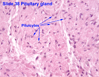
3. PITUICITO: is a glial cell of the posterior pituitary. [1]
They generally stain dark purple, and are among the easiest structures to identify in the region (pars nervosa of the posterior pituitary). [2][3] They are similar to the astrocytes/glial cells of the CNS.
Uniones comunicantes que mantienen composicion ionica extracelular
OXT contractilidad del miometrio
ADH nivel hidrico y osmotico

Tiroides

Foliculos: The thyroid is composed of spherical follicles that selectively absorb iodine (as iodide ions, I-) from the blood for production of thyroid hormones, but also for storage iodine in thyroglobulin, in fact iodine is necessary for other important iodine-concentrating organs as breast, stomach, salivary glands, thymus etc. (see iodine in biology).
Twenty-five percent of all the body's iodide ions are in the thyroid gland. Inside the follicles, colloid serve as a reservoir of materials for thyroid hormone production and, to a lesser extent, act as a reservoir for the hormones themselves. Colloid is rich in a protein called thyroglobulin.
1. EPITELIO CUBICO SIMPLE
2. MEMBRANA BASAL
3. CONECTIVO INTER FOLICULAR4. ENDOTELIO
5. COLOIDE
Celulas Foliculares: are cells in the thyroid gland that produce and secrete:
-thyroxine(T4)
-triiodothyronine (T3).
-thyroxine(T4)
-triiodothyronine (T3).
The main function of the thyroid gland is to take iodine, found in many foods, and convert it into thyroid hormones: thyroxine (T4) and triiodothyronine (T3).Thyroid cells are the only cells in the body which can absorb iodine. These cells combine iodine and the amino acid tyrosine to make T3 and T4. T3 and T4 are then released into the blood stream and are transported throughout the body where they control metabolism (which is the conversion of oxygen and calories to energy). Every cell in the body depends upon thyroid hormones for regulation of their metabolism. The normal thyroid gland produces about 80% T4 and about 20% T3, however, T3 is about four times as potent as T4. They are simple cuboidal epithelium and are arranged in spherical follicles surrounding colloid.
They have thyrotropin receptors on their surface, which respond to thyroid-stimulating hormone.
Parafoliculares: are cells in the thyroid that produce and secrete calcitonin. They are located adjacent to the thyroid follicles and reside in the connective tissue. These cells are large and have a pale stain compared with the follicular cells or colloid. In teleost and avian species these cells occupy a structure outside of the thyroid gland named the ultimobranchial body.
In a series of elegant experiments, Nicole LeDouarin (ref), transplanted neural crest cells from quail, with unique and easily identified nuclei, into the neural crest of ____. She subsequently demonstrated the presence of cells with quail nuclei populating the ultimobranchial body and concluded that C cells migrate during embryologic development from the neural crest.
Embryologically, they associate with the ultimobranchial body, which itself is a ventral derivative of the fourth (or fifth[1]) pharyngeal pouch. Parafollicular cells themselves are derived from Neural Crest cells. They are not numerous in the thyroid and are typically situated basally in the epithelium, without direct contact with the follicular lumen. They are always situated within the basement membrane, which surrounds the entire follicle.
When parafollicular cells become cancerous, they lead to medullary carcinoma of the thyroid.